
DUAL-ENERGY CT FOR GOUT

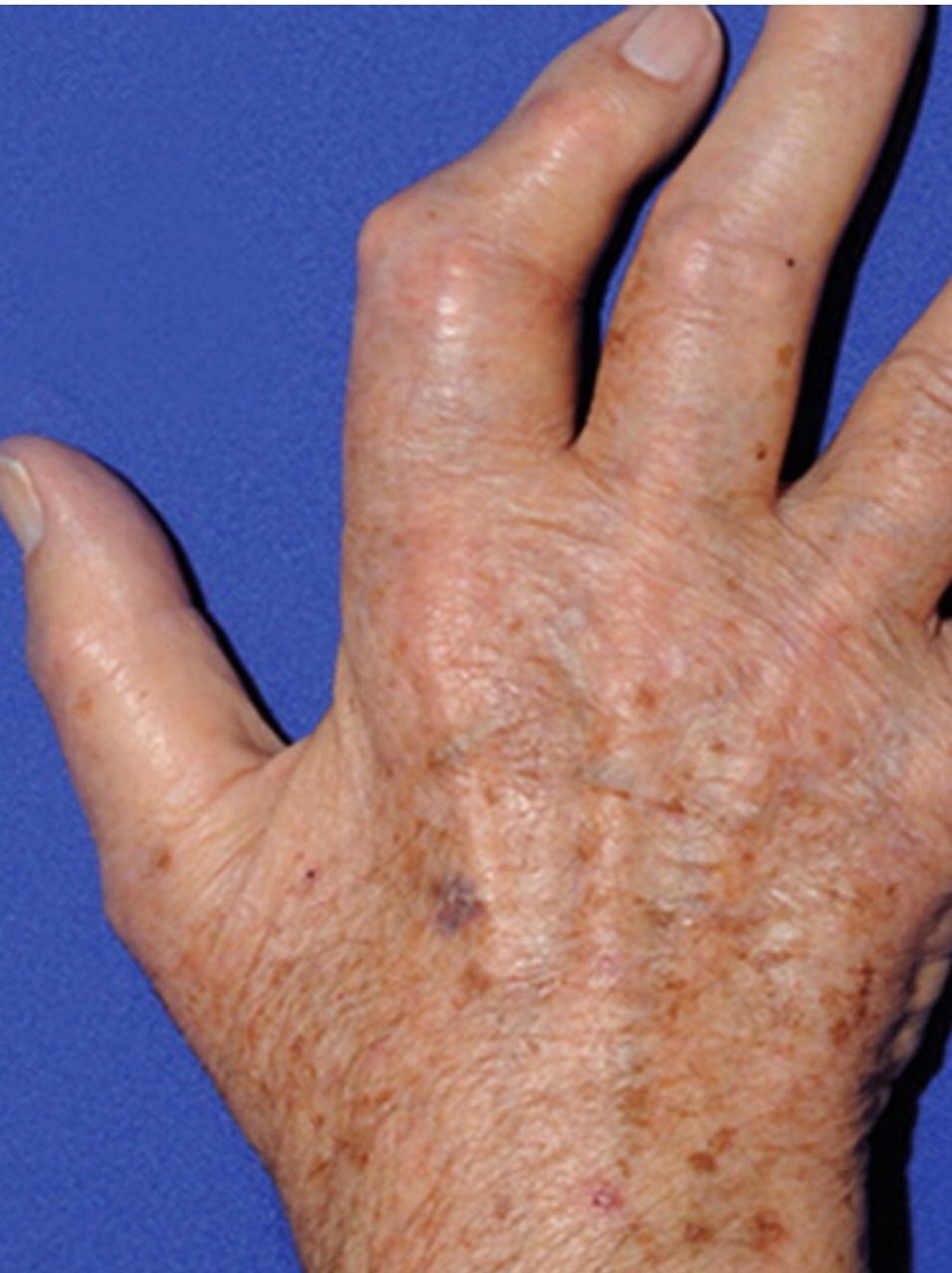
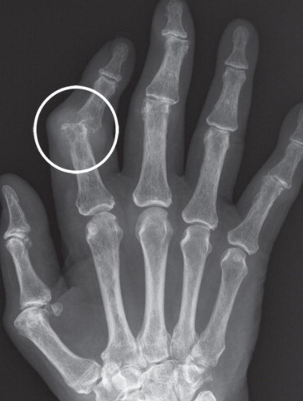

Acute gouty arthritis of the first metatarsophalangeal joint, termed “podagra,” was first identified by Egyptians in 2640 B.C. and continues to be a medical health problem today. The hallmark of gout is hyperuricemia with subsequent deposition of monosodium urate (MSU) crystals, which leads to inflammation and symptoms. Gout commonly involves specific joints and anatomic structures, and knowledge of these sites and imaging appearances are clues to the correct diagnosis.
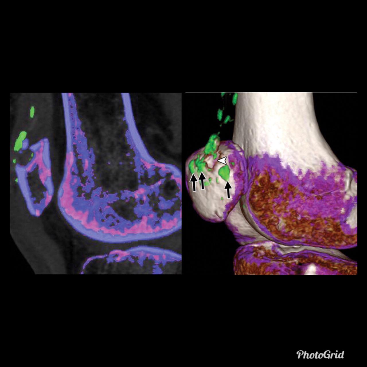
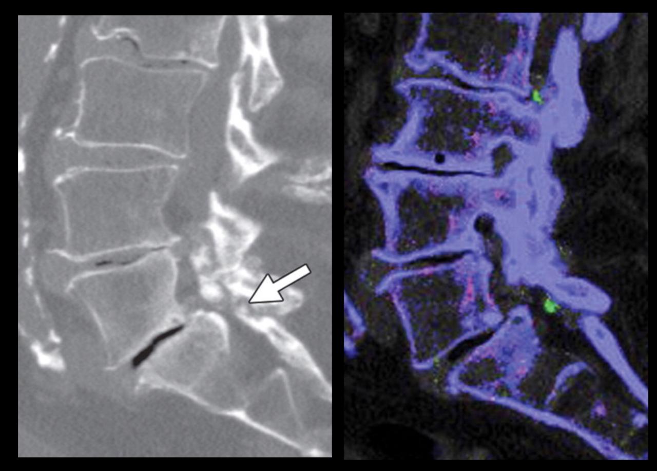
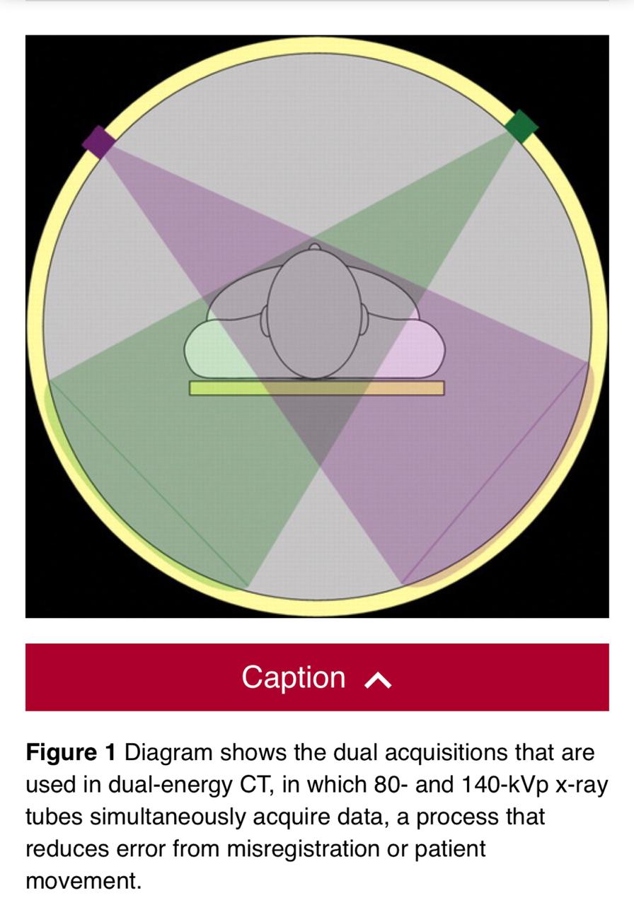
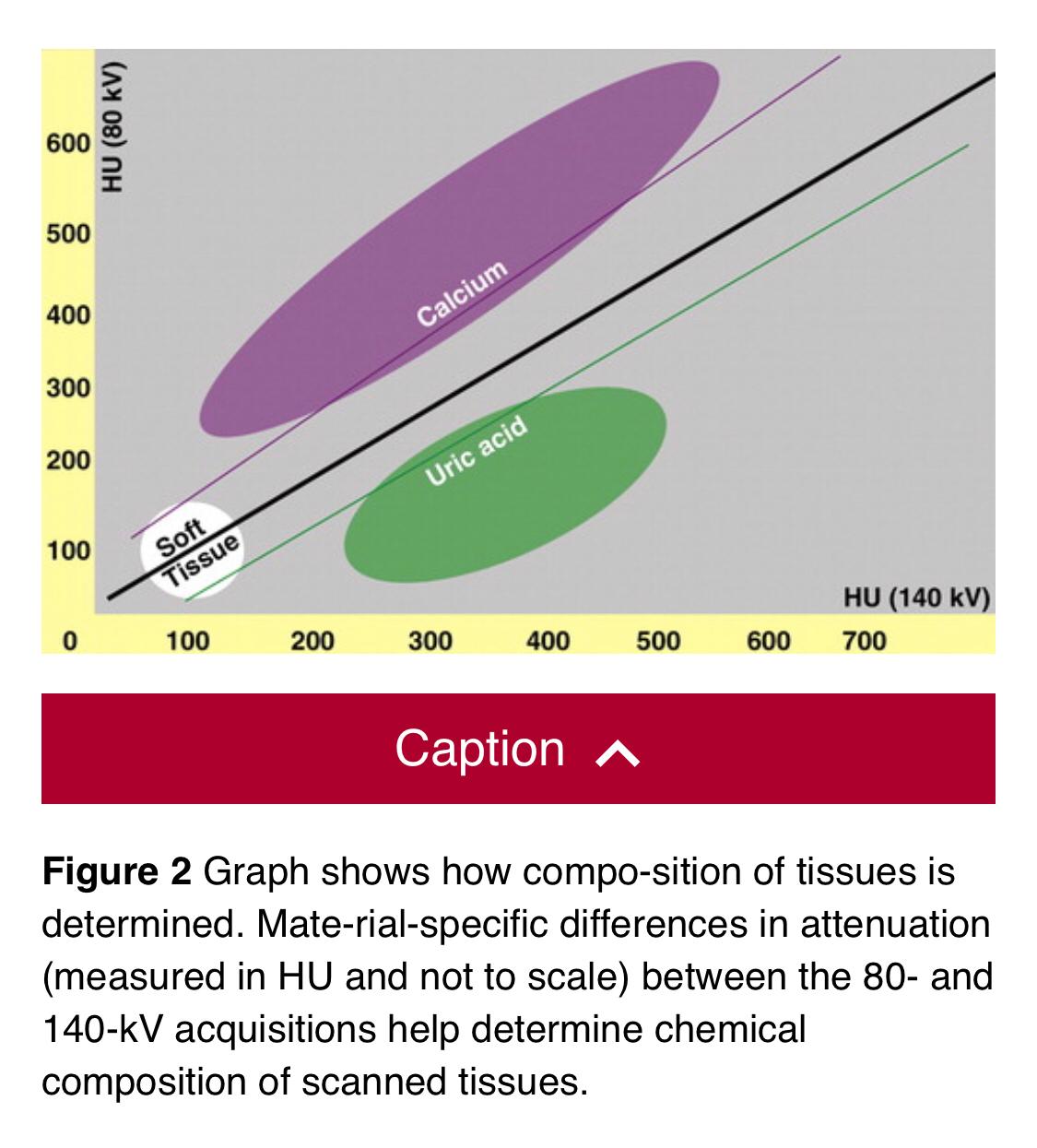
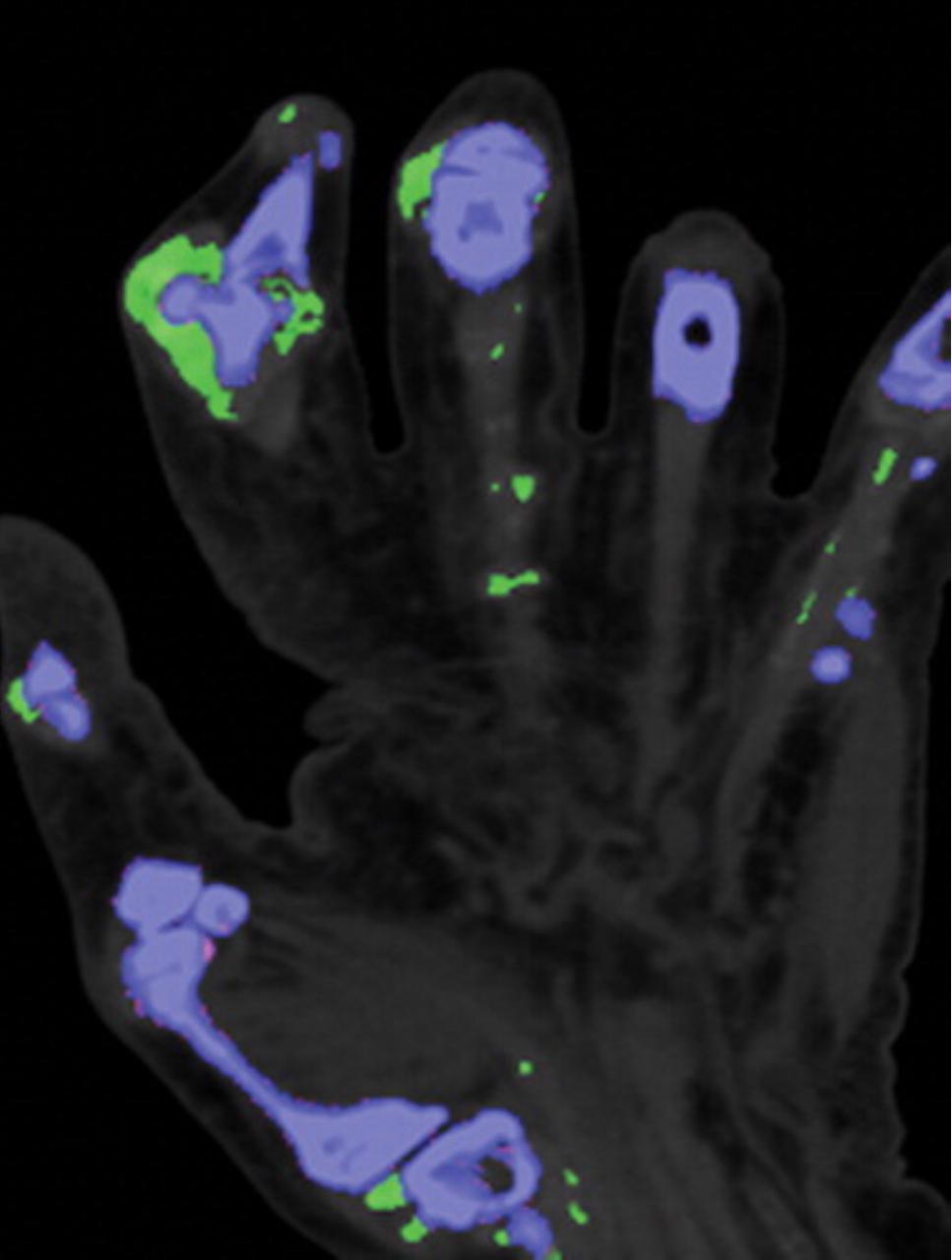
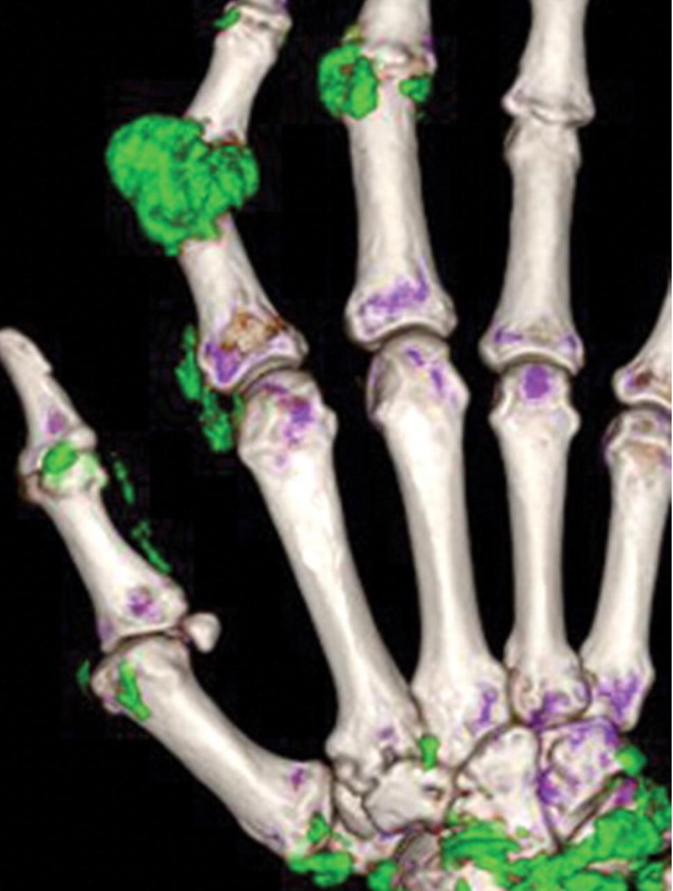
Although diagnosing gout generally is straightforward, atypical disease may present a challenge if it is associated with unusual symptoms or sites, discordant serum urate level, or mimics of gout. Dual-energy computed tomography (CT) may be used to differentiate urate crystals from calcium by using specific attenuation characteristics, which may help diagnose gout. In patients with known tophaceous gout, dual-energy CT may be used for serial volumetric quantification of subclinical tophi to evaluate response to treatment.
Dual-energy CT can quantitatively identify monosodium urate crystal deposits with high sensitivity and specificity within joints, tendons, and periarticular soft tissues.
Given the utility of dual-energy CT in challenging cases and its ability to provide an objective outcomes measure in patients with tophaceous gout, dual-energy CT promises to be a unique and clinically relevant modality in the diagnosis and management of gout.